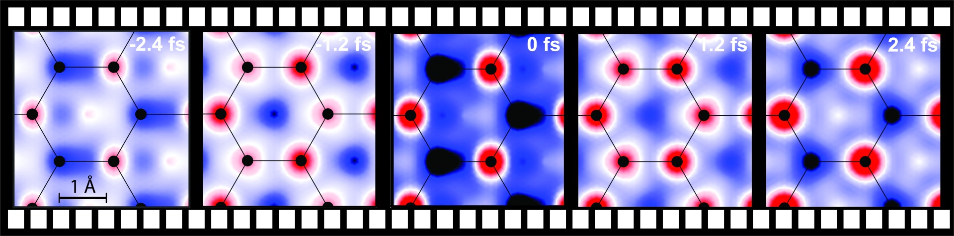
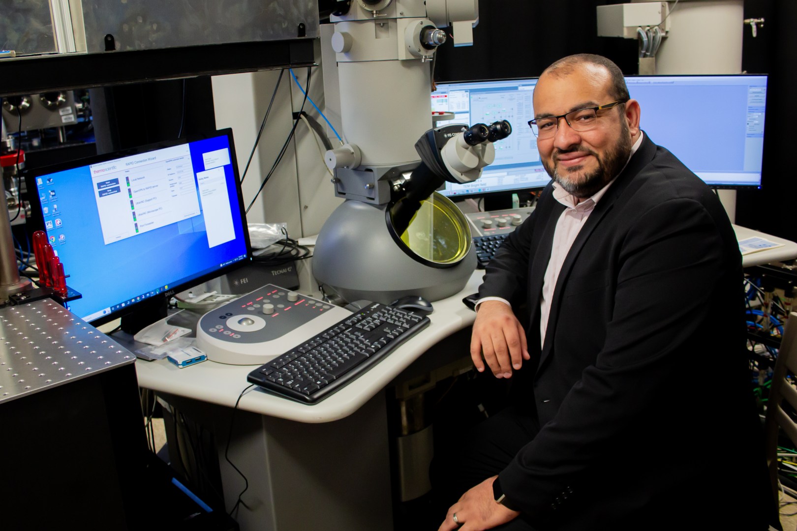
Left: A conceptual representation of Attomicroscopy; Right: The device and its surrounding equipment.
Mohammad Hasan/University of Arizona
Researchers at the University of Arizona have created the world’s fastest electron microscope, a remarkable device capable of capturing freeze-frame images of moving electrons.
They anticipate that this innovation will lead to significant breakthroughs in fields such as physics, chemistry, bioengineering, materials science, and beyond.
“This new tool has the highest temporal resolution to freeze time and see electron motion in action. This Attomicroscopy electron imaging builds a strong bridge to convert scientific findings to engineering applications,” Mohammed Hassan, associate professor of physics and optical sciences, told Interesting Engineering (IE).
“With this microscope, we hope the scientific community can understand the quantum physics behind how an electron behaves and how an electron moves.”
How does electron microscopy work?
Ultrafast electron microscopes, first developed in the 2000s, utilize lasers to generate pulsed electron beams, significantly enhancing the temporal resolution—the ability to observe changes in a sample over time.
Unlike traditional microscopes, where image quality depends on the camera’s shutter speed, the resolution in these advanced transmission electron microscopes is determined by the duration of the electron pulses.
The principle is straightforward: the faster the pulse, the sharper the image.
Earlier ultrafast electron microscopes operated by emitting a series of electron pulses at speeds measured in attoseconds—one quintillionth of a second. These pulses created sequential images, much like frames in a movie, but the rapid changes and reactions within an electron between those frames remained elusive.
For the first time, University of Arizona researchers generated a single attosecond electron pulse to capture an electron in a fixed state, matching the speed at which electrons move. This breakthrough significantly enhances the microscope’s temporal resolution, akin to a high-speed camera capturing moments that would otherwise be invisible.
Expanding Nobel Prize-winning theories
Hassan and his team built upon the Nobel Prize-winning work of Pierre Agostini, Ferenc Krausz, and Anne L’Huillier, who were awarded the Nobel Prize in Physics in 2023 for generating the first extreme ultraviolet radiation pulse short enough to be measured in attoseconds.
“The great impact of these three pioneer scientists allowed us to trace electron motion in real-time using attosecond light pulses. Our work builds upon and extends the foundation laid by their efforts by generating the first attosecond electron pulses, adding the capability of seeing electron motion in space and time simultaneously,” Hasan tells IE.
Imaging moving electrons
Leveraging this groundbreaking achievement, the University of Arizona researchers developed a cutting-edge microscope that uses a powerful laser split into two components: a highly rapid electron pulse and two ultra-short light pulses.
The first light pulse, known as the “pump pulse,” energizes the sample, causing electrons to move or undergo other rapid changes. The second pulse, referred to as the “optical gating pulse,” acts as a gate by creating a brief time window during which a single attosecond electron pulse is generated. The timing of this gating pulse determines the resolution of the resulting image.
By meticulously synchronizing these two pulses, researchers can precisely control when the electron pulses interact with the sample, allowing them to capture and observe ultrafast processes at the atomic level.
Attomicroscopy and material, chemical, and biological sciences
When asked about the significance of this major milestone and what it would allow us to see that we couldn’t previously, Hasan says, “Attomicroscopy connects the material structure morphology with its electron dynamics, giving us the possibility to image and control the electron currents and develop laser field-driven electronics a million times faster than the current electrons, and it can be as small as a few nanometers.”
“The impact of Attomicroscopy is not only on engineering applications but also can extend to chemistry and biology. Seeing the electron motion with Attomicroscopy can be extended to see the chemical boding breaking and formation,” he adds.
“This new capability would allow us to realize the long-anticipated dreams of chemists to control the chemical reactions in situ. Hence, we can create new molecules and revolutionize the drug discovery research area.”
Regarding applications in biological science, the team lead explains, “Attomicroscopy would also benefit from the great advancement in cryo-TEM (which received the Nobel Prize in 2017) to see the electron motion in biological molecules and samples. Imagine if we could see how the electrons move and control them on demand to confirm the DNA 3D structure. This, indeed, will open the door for more scientific and technological breakthroughs.”
Hasan with his team’s innovation.
How does the technology translate to application-based science?
So, what does this mean for advances in usable technology? Hasan says, “A perfect example of that is we used the insights we learned from imaging the electron motion in graphene by Attomicroscopy in this study to develop attosecond current switches and ultrafast graphene-based optoelectronics in another work currently under publication.”
This would mean the development of ultrafast, nearly six orders of magnitude faster, optical transistors, lightwave electronics, and optical quantum computers.
RECOMMENDED ARTICLES
When asked about what this development means for the so-called ‘mysterious’ nature of quantum physics and why is it necessary to see an electron in motion, Hasan explains, “This is a very important question. We are trying to understand the quantum behavior of electron motion from the static perspective.”
“Attomicroscopy would open a new gate in time for us to see and understand the quantum behavior of electrons in real time and space. Attomicroscopy could potentially be the quantum camera to film this mysterious world,” he concludes.
Srishti Gupta Srishti studied English literature at the University of Delhi and has since then realized it's not her cup of tea. She has been an editor in every space and content type imaginable, from children's books to journal articles. She enjoys popular culture, reading contemporary fiction and nonfiction, crafts, and spending time with her cats. With a keen interest in science, Srishti is particularly drawn to beats covering medicine, sustainability, gene studies, and anything biology-related.


0